


71 A, Suriya Hospital, Puliyangudi, Tenkasi - 627855
An USG scan (ultrasonography) is an imaging examination that uses high-frequency sound waves to create pictures of structures inside the body. Depending on the area being examined, an ultrasound may help a doctor assess organs, soft tissues, fluid-filled structures or pregnancy-related development.
An ultrasound is usually recommended based on a person's symptoms, clinical findings, pregnancy status, or a specific medical question. The type of scan and preparation required can therefore differ from one patient to another. At Surya Hospital, Puliyangudi, patients can discuss their requirements with the healthcare team to understand which diagnostic evaluation may be appropriate.
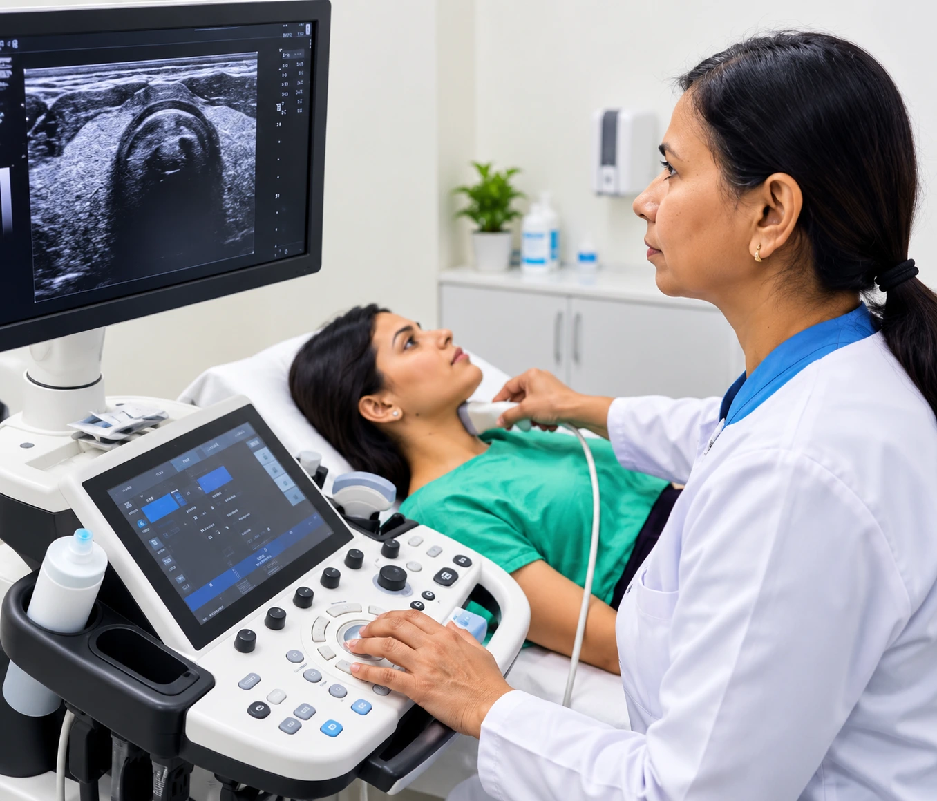
A USG scan is a non-invasive diagnostic imaging test that produces real-time images using sound waves rather than ionizing radiation. A trained healthcare professional places an ultrasound transducer over the area being examined, often using a gel to help the sound waves travel effectively.
The resulting images can help the treating doctor evaluate particular anatomical structures and look for findings that may require further assessment. Ultrasound is an imaging tool rather than a diagnosis by itself, so the scan findings need to be interpreted in the context of the patient's symptoms and clinical examination.
An ultrasound may be requested when a doctor needs more information about a particular symptom, physical finding or suspected condition. It can also be used in pregnancy to assess relevant aspects of fetal development and the uterus.
The reason for the examination matters because one type of ultrasound is not suitable for every diagnostic question. The body area being examined, the patient's age, symptoms, pregnancy status and the information the doctor needs can all influence the choice of scan.
The exact purpose should be explained by the treating doctor or healthcare professional who requested the examination.
Ultrasound can be useful for examining many structures because it provides real-time imaging without using X-rays. Depending on the type of examination, it may help evaluate organs, tissues, fluid collections or other structures within the area being scanned.
However, ultrasound has limitations. The quality and usefulness of the images can be affected by factors such as the body area being examined, the patient's anatomy and the nature of the condition. Some medical problems may require a different imaging test or additional evaluation.
This is why the appropriate scan should be selected according to the clinical question rather than simply choosing an ultrasound because it is readily available.
“USG scan” is a broad term rather than one single examination. Ultrasound examinations can differ according to the body part being assessed and the purpose of the investigation.
For example, a doctor may request an ultrasound examination of the abdomen, pelvis or another specific region when clinically appropriate. Pregnancy-related ultrasound examinations also have their own indications and may be performed at different stages of pregnancy.
The specific ultrasound examinations available at Surya Hospital should be confirmed with the hospital before booking, as the service offered can depend on the diagnostic requirement and available facilities.

Yes. Ultrasound uses sound waves to produce images, while X-rays and CT scans use ionizing radiation. Ultrasound also provides real-time imaging and is particularly useful for certain soft-tissue and fluid-related assessments.
However, the tests are not interchangeable. Each imaging method has different strengths and limitations. A doctor may choose an ultrasound, another imaging examination, or additional testing depending on what needs to be evaluated.
Ultrasound does not use ionizing radiation and has been widely used for diagnostic imaging, including during pregnancy when medically indicated.
Like any medical examination, it should be performed for an appropriate clinical purpose. The type of ultrasound, its timing and whether further testing is needed should be determined according to the patient's individual circumstances.
An ultrasound may be appropriate for patients with particular symptoms, clinical findings or diagnostic concerns when their doctor considers it useful. It may also be recommended during pregnancy for specific assessment purposes.
It is important not to assume that every symptom requires an ultrasound. The need for imaging depends on the clinical situation and the question the doctor is trying to answer.
Before scheduling an ultrasound, patients should check whether any preparation is required. Preparation can vary significantly depending on the body area being examined.
For some examinations, the healthcare provider may give instructions regarding food or fluid intake, while another type of scan may require different preparation. Following the instructions provided for the specific examination can help ensure that the scan is performed under the appropriate conditions.
If you have been advised to undergo an ultrasound, carrying your doctor's prescription, previous scan reports and relevant medical records can also help the healthcare team understand the reason for the examination.
An ultrasound is most useful when it is connected to a clear clinical question. The scan itself provides images; the significance of those findings depends on the patient's symptoms, examination and other available medical information.
If your doctor has recommended a USG scan, understanding why it was requested, what area will be examined, whether preparation is required and what happens after the scan can make the diagnostic process clearer and less stressful.
Preparation for an ultrasound scan depends on the type of examination and the body area being assessed. Some scans may require you to avoid food or drink for a specific period, while others may require a comfortably full bladder. Not every ultrasound needs special preparation.
Before your appointment, follow the instructions provided by the healthcare team. If you are unsure whether you should eat, drink water, take your regular medicines, or arrive with a full bladder, confirm the requirements before the examination rather than assuming the same preparation applies to every USG scan.
The preparation instructions are part of the diagnostic process because suitable conditions can help the healthcare professional obtain appropriate images.
Before the examination begins, the healthcare professional may confirm the reason for the scan and the area that needs to be assessed. You may be asked about relevant symptoms, previous investigations or other information connected with the examination.
Depending on the type of ultrasound, you may need to change position, expose the area being examined, or follow a specific preparation instruction. If you are having an ultrasound for a particular medical concern, providing relevant previous reports can help place the examination in context.
The scan itself is generally focused on obtaining images of the area requested by the doctor. It is not intended to replace the clinical assessment that led to the investigation.
During an ultrasound examination, you will usually lie in a suitable position so the area being examined can be accessed. A small amount of ultrasound gel may be applied to the skin, and a handheld device called a transducer is moved over the area.
The transducer sends and receives sound waves, which are processed to create images on a monitor. The healthcare professional may move the transducer across different parts of the area being examined or ask you to change position when necessary.
Depending on the examination, you may be asked to remain still or briefly adjust your position. The duration and process can vary according to what needs to be assessed.
A standard ultrasound examination is generally well tolerated because the transducer is placed over the skin rather than requiring an incision. You may feel pressure as the device is moved over the area being examined.
If the area being scanned is already painful or tender, the pressure may feel uncomfortable. Certain examinations may also involve specific techniques depending on the body area and clinical purpose. If you experience significant discomfort during the examination, inform the healthcare professional performing the scan.
The time required for a USG scan can vary depending on the type of examination, the area being assessed and the images that need to be obtained.
A straightforward examination may be relatively brief, while a more detailed assessment can take longer. Preparation requirements can also affect the overall appointment time.
Rather than relying on a fixed duration, patients should follow the instructions given for their particular examination and allow sufficient time for registration, preparation and the scan itself.
Obtaining useful ultrasound images depends on several factors. The body area being examined, the nature of the finding and the patient's individual anatomy can all influence what can be visualised.
Gas within the digestive tract, for example, can interfere with the passage of ultrasound waves in some examinations. This is one reason why preparation instructions may be provided for particular scans.
The quality of the examination can also depend on the specific diagnostic question and whether ultrasound is the most suitable imaging method for that concern. If the images do not provide enough information, your doctor may consider whether another investigation is appropriate.
Once the required images have been obtained, the examination is completed and you can generally continue with your activities according to the instructions provided.
The images are assessed as part of the reporting process. The significance of any finding should be considered alongside your symptoms, examination and medical history rather than interpreting an isolated image without clinical context.
If the scan was requested by a doctor, the next step may be to discuss the report with that doctor. Depending on the findings, your doctor may recommend observation, further investigation, treatment or another appropriate follow-up plan.
Not always. Ultrasound can provide important information, but it has diagnostic limitations and is not the best imaging method for every condition.
A scan may identify a finding that requires further assessment, or the images may not provide enough information to explain a patient's symptoms. In such situations, additional clinical evaluation or another imaging test may be considered.
This does not necessarily mean that something serious has been found. It means that different diagnostic tools answer different medical questions, and your doctor decides what information is needed next.
A doctor may request another investigation when the ultrasound findings are unclear, when a more detailed assessment is needed, or when the suspected condition is better evaluated using another imaging method.
The choice can depend on the body area, suspected condition, previous findings and the patient's individual circumstances. Additional imaging should therefore not be viewed as a failure of the ultrasound examination; it may simply provide information that ultrasound cannot adequately supply.
Your doctor can explain why another test is being considered and how it relates to the original USG findings.
An ultrasound report contains medical observations made from the examination. Some terms may be difficult to understand without clinical context, particularly when they describe measurements, anatomical structures or findings that require comparison with symptoms and previous investigations.
Rather than trying to diagnose yourself from the report, discuss the findings with the doctor who requested the scan. The doctor can explain what the findings mean for your particular situation and whether any follow-up is necessary.
If you have previous ultrasound or other imaging reports, keeping them available can also help with comparison when your doctor considers whether a finding has changed over time.
An abnormal or unexpected finding does not automatically mean that you have a serious disease. Ultrasound findings need to be interpreted according to the patient's age, symptoms, medical history and the characteristics of the finding.
Your doctor may recommend further assessment, repeat imaging, laboratory testing, specialist consultation or treatment depending on what the examination shows. The appropriate next step is therefore based on the complete clinical picture, rather than the scan result alone.
These practical steps can help you understand the purpose of the examination and what should happen after it.
Follow-up depends on why the ultrasound was performed and what the examination shows. Some scans may simply provide information for the treating doctor, while others may lead to additional testing or a planned review.
If your doctor has specifically asked you to return with the report, follow the recommended appointment schedule. If your symptoms continue or change after the examination, inform your healthcare provider rather than assuming that a previous scan rules out the need for further medical assessment.
Choosing where to have an ultrasound scan is not only about finding a diagnostic facility. Patients also need a clear understanding of why the scan has been advised, what preparation is required, what the examination involves and what should happen after the report is available.
At Surya Hospital, USG scans form part of the diagnostic support available to patients who have been advised ultrasound evaluation. The appropriate examination depends on the clinical requirement and the area that needs to be assessed. Patients can discuss their scan requirement with the hospital team and follow the preparation instructions provided for their examination.
Surya Hospital is located at:
71 A, Surya Hospital, 5th West Street, Karpaga Veethi, Opposite to T.N.S.T.C Bus
Depot, Puliyangudi, Tenkasi - 627855
Patients from Puliyangudi and surrounding areas of Tenkasi district can consider the hospital when they have been advised a USG scan as part of their medical evaluation.
When selecting an ultrasound facility, patients should look beyond location alone. The most appropriate choice depends on the reason for the examination and whether the facility can support the required diagnostic process.
The goal should not simply be to obtain an ultrasound image, but to ensure that the examination contributes appropriately to the overall diagnostic process.
For patients whose diagnostic evaluation relates to a surgical concern, the treating doctor may need to interpret the findings alongside the clinical examination and other available information.
Dr. M. Sivasubramanian, Managing Director of Surya Hospital, is a General & Laparoscopic Surgeon with qualifications including M.S., D.MAS., F.MAS., FIAGES, FALS and F.ART.
For surgical conditions, an ultrasound finding may be one component of the assessment rather than the sole basis for deciding treatment. The doctor can determine whether further evaluation or management is appropriate based on the patient's individual circumstances.
For women undergoing ultrasound as part of obstetric or gynecological evaluation, consultation should be with the appropriate Obstetrics & Gynecology professional.
A USG scan may be requested alongside other investigations depending on the patient's symptoms and clinical needs. The appropriate combination of tests should be determined by the treating healthcare professional rather than selected solely on the basis of symptoms.
Patients may also require consultation related to the hospital's other healthcare services, including Laparoscopic & General Surgery and Obstetrics & Gynecology, depending on the reason for their diagnostic evaluation.
If an ultrasound identifies a finding that requires clinical attention, the next step can be discussed with the appropriate doctor.
71 A, Surya Hospital, 5th West Street, Karpaga Veethi, Opposite to T.N.S.T.C Bus Depot, Puliyangudi, Tenkasi - 627855
It depends on the type of ultrasound. Some examinations may require a comfortably full bladder, while others do not. Follow the specific preparation instructions provided for your scan rather than drinking large amounts of water without guidance.
Some abdominal examinations may require fasting, but fasting is not necessary for every USG scan. The preparation depends on the body area and purpose of the examination. Confirm the instructions for your particular scan before your appointment.
Medication instructions can vary depending on your medical circumstances and the type of examination. Do not stop prescribed medicines on your own. If fasting has been advised, ask the healthcare team whether any regular medication should be taken before the scan.
Ultrasound is commonly used during pregnancy when clinically indicated because it uses sound waves rather than ionizing radiation. The type and timing of an examination should be determined by the treating obstetrician according to the pregnancy and clinical requirement.
No. Ultrasound is useful for evaluating many organs, tissues and conditions, but it cannot detect or conclusively diagnose every disease. Depending on the clinical question, your doctor may recommend another investigation or additional testing.
A repeat or additional scan may be recommended when further assessment is needed, when the initial images are inconclusive, or when follow-up comparison is clinically useful. The recommendation depends on the findings and your individual medical situation.
If your examination required fasting, you can generally resume eating after the examination unless your healthcare provider has given different instructions. If you did not need to fast, there may be no food-related restriction after the scan.
Yes, when they are relevant to the current examination. Previous reports or images can provide useful background and may help your treating doctor compare findings when comparison is appropriate.
The report can describe the findings, but interpreting their medical significance requires clinical context. A doctor can relate the report to your symptoms, examination and medical history and explain whether further evaluation is required.
A normal ultrasound does not necessarily explain every symptom or exclude every possible condition. If symptoms continue, worsen or change, consult your treating doctor. Further clinical assessment or another investigation may be considered depending on the situation.
If your doctor has advised an ultrasound examination, understanding the required preparation and bringing relevant medical information can make the appointment easier. The type of USG scan should be based on the clinical requirement, and any preparation instructions should be followed carefully.
For patients looking for a USG scan in Puliyangudi, Tenkasi, Surya Hospital provides a local option for diagnostic evaluation. Speak with the hospital team regarding your scan requirement, preparation instructions and appointment details.